
Global Health
Focusing on abnormal fundus examination results
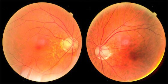
Fundus examination, also called ophthalmoscopy or retinal examination, is a test used to detect eye disorders, injuries and diseases. Advanced practice nurses working in primary care and emergency departments should develop and improve their fundus skills as it may possibly be considered one of the harder procedures performed during each routine physical examinations and emergency care assessments. Use our Fundus Examination pocket card to familiarize yourself with the anatomy, equipment and steps involved in performing a fundus examination.
Abnormal results (Stanford Medicine, n.d.; Schneiderman, 1990)
Once you are feeling comfortable examining the fundus and develop into accustomed to the traditional anatomic landmarks, begin to enhance your skills in detecting abnormal findings. Please listen to the next:
- : swelling of the optic disc with swollen veins and retinal hemorrhages. This is a medical emergency and an indication of increased intracranial pressure, which could also be brought on by a tumor, hemorrhage, or thrombosis.
- : bleeding because of ruptured deep capillaries or large vessels that happens in endocarditis, pernicious anemia, diabetic retinopathy, leukemia, and subarachnoid hemorrhage.
- : the optic disc will appear raised and covered with cotton wool spots (fluffy gray-white or yellow spots with blurred edges), which could also be visible in cases of chronic diseases akin to hypertension, diabetes, HIV, severe anemia or thrombocytopenia, hypercoagulable states, tissue total disorders and viruses.
- : Chronic hypertension stiffens and thickens arteries. Where arteries and veins cross, the arteries will “cut into and displace” the veins. This is seen in chronic vascular disease.
- : brought on by platelet, fibrin or cholesterol emboli that cause a gray area to form above the plaque.
- : inflammation of the attention’s uvea (iris, ciliary body and choroid) which can involve the retina, retinal vessels, vitreous humor, optic nerve head and sclera. It appears as a yellow-white lesion with blurred edges, described as a “spotlight in the fog”. It might be brought on by toxoplasmosis, cytomegalovirus (CMV), herpes simplex, rubella, West Nile virus, HIV-related eye diseases, tuberculosis, syphilis, and fungal infections, amongst others (Geetha and Tripathy, 2022).
- could also be brought on by corneal scarring, cataracts, or vitreous hemorrhage.
- on the optic disc are an indication of proliferative diabetic retinopathy.
- may indicate glaucoma or high myopia.
- are yellow deposits of lipids and proteins under the retina that naturally form with age. The presence of numerous detected drusen is an early sign of age-related macular degeneration (AMD). Late AMD is related to rapid vision loss.
- observed on prolonged examination may indicate uveal melanoma, essentially the most common primary intraocular tumor in adults. Most develop within the choroid, ciliary body, and iris (Tarlan and Kiratli, 2016).
- best viewed with dilated pupils on indirect fundoscopy. The unit could have a dune-like appearance and the volleyball will appear mobile. Often you possibly can see the opening that caused the detachment.
Good luck specializing in your fundoscopy skills! Continue to practice and develop a standardized approach to this common procedure to detect chronic diseases and life-threatening emergencies.
Geetha, R. and Tripathy, K. (2022, August 22). Inflammation of the choroid and retina. . https://www.ncbi.nlm.nih.gov/books/NBK551705/#
Schneiderman, H. (1990). Fundus examination. Clinical methods: history, physical and laboratory examinations. 3R&D Editing.
https://www.ncbi.nlm.nih.gov/books/NBK221/
Stanford Medicine. (n.d.) Introduction to fundus examination/ophthalmoscopy. . https://stanfordmedicine25.stanford.edu/the25/fundoscopic.html
Tarlan, B. and Kıratlı, H. (2016). Uveal melanoma: current trends in diagnosis and treatment. , (3), 123–137. https://doi.org/10.4274/tjo.37431

What does “stronger than chemicals” mean?

Beyond community services: common cleanliness gaps in patient care

The missing decade: Nursing informatics could shape the longer term of menopause care

Hidden assumptions that put nurses prone to sexual harassment

Global Fund Board Elects New Chairman and Vice-Chairman – Press Releases

Global Fund Welcomes Indonesia’s $10 Million Commitment for Eighth Replenishment – Updates

5 books that may help at work at work

The Global Fund opens up the potential of private sector investment – updates

Fast and healthy advice on preparing meals for busy nurses

Maintenance of the nursing engine – each day nurse

Safety within the workplace as an ethical imperative in nursing

A cultural approach to the treatment of neonatal pain
-
 Well-Being1 year ago
5 books that may help at work at work
-
 Global Health1 year ago
The Global Fund opens up the potential of private sector investment – updates
-
 Well-Being1 year ago
Fast and healthy advice on preparing meals for busy nurses
-
 Well-Being1 year ago
Maintenance of the nursing engine – each day nurse
-
 Best Practice1 year ago
Safety within the workplace as an ethical imperative in nursing
-
 Best Practice1 year ago
A cultural approach to the treatment of neonatal pain
-

 Well-Being1 year ago
How to get the standard of sleep for higher mental health
-

 Education1 year ago
AI for teachers – Nursing Education Network